


Crooked legs and Inflamed Growth Plates in Foals – November 2020
Crooked legs and Inflamed Growth Plates in Foals – November 2020
November 1, 2020
Barbara Hunter DVM MS DACVS-LA
Angular limb deformities or ‘crooked legs’ in foals can result from a number of factors. Needless to say, treatment is directed by the underlying cause. Fortunately in many cases, treatment is economic as it consists of rest and dietary management. In some cases, however, treatment requires surgery.
The most straightforward explanation of angular limb deformities is a bend in the foal’s leg, either to the outside or inside of centerline. In newborn foals, the most common reason for this to occur is laxity in the ligaments supporting its joints. This typically responds well to confinement with small periods of controlled exercise (eg: 1-2 hours of turnout) for the first 2-3 weeks of life. In cases were the foal appears immature or has been born prematurely, angular limb deformities can be present due to ‘weak’ or underdeveloped bones within the knees and hocks. These cases are vital to recognize and treat as soon as possible, as delayed treatment can lead to permanent malformation of the limb. The first step in recognition is having a veterinarian examine the foal and take radiographs of the affected joint to ensure that the bones are properly formed.
In slightly older foals (1-4 months old), legs that had started out straight can slowly take on a bent appearance. Most commonly the bend starts at the level of a growth plate just above a joint and is a result of one side of the growth plate growing faster than the other. If this is caught early, the bend is often easily corrected with some simple management changes: corrective farriery, moderate confinement and minimizing supplemental feeding. In cases where the bend is severe, or the foal is near the age when the growth plate closes, rapid correction is essential. Once the growth plate closes, the opportunity for the slower growing side of the limb to ‘catch up’ and straighten the leg has been lost. These are the types of cases that require surgical correction. Particularly in cases of fetlock angular limb deformity, where the affected growth plate closes between 12 and 16 weeks of age, correction of the deformities early in their development is important to achieve a good result.
Physitis or inflammation of growth plates does not cause deviation of a limb, but it can cause lameness and unsightly swelling of growth plates. This is most commonly seen in weanling to yearling aged horses, particularly during periods of rapid growth. Treatment is geared toward slowing growth and decreasing pressure on the limbs. Affected horses should be confined and controlled exercise minimized. It is important to decrease their plane of nutrition both to slow growth and encourage some weight loss. Finally, treatment with systemic anti-inflammatories, such as phenylbutazone, is important in the short term to decrease the inflammation within the growth plate.
Equine Osteochondrosis Dissecans
August 26, 2020
Equine Osteochondrosis Dissecans – September 2020
B Hunter DVM MS MANZCVS DACVS-LA
Osteochondrosis dissecans (OCD) is a disease affecting joint cartilage and bone in juvenile horses that has a serious economic impact on the equine industry. The disease has been heavily researched for decades. Despite this, a clear understanding of the underlying causes still eludes experts. The following is an explanation of the basic underlying events, clinical signs, diagnosis and treatment of the disease, as well as discussion of the most important factors currently thought to influence the development of this multifactorial disease.
The traditional definition of OCD is that it is a failure in a process called endochondral ossification in the neonate. While more current research shows that that is not precisely true, understanding this relatively straightforward process in normal development does give one a base upon which to build more complex ideas.
When a foal is formed in utero, its bones start out as a cartilage template. This template then ossifies or mineralizes into bone in the process known as endochondral ossification. The mineralization process starts from specific centers, known as ossification centers, within the bone and these centers are in predictable locations. In long bones, the main part of the bone (diaphysis) has an ossification center in the middle and mineralization moves toward both ends of the bone. At the end of each bone are separate centers of ossification known as epiphyses (Figure 1). These epiphyses are important as they are the part of the bone that enter joints and are affected by OCD. When a foal is born, endochondral ossification is complete within the long part of the bone, but not at the epiphyses. These areas still have thick layers of cartilage that need to develop into bone. That is why the boney aspect of long bone ends is rounder and smaller in neonates than adults. Disruption to this cartilage as it ossifies is what leads to OCD lesions. The tricky part is understanding that disruption is instigated by a combination of factors. The most influential of these factors varies by breed, environment and the joint affected.
The primary clinical sign of OCD is effusion or extra fluid within a joint in a juvenile horse. Most commonly this effusion develops when a horse is first started into work, thus the age at which clinical signs are seen varies between breeds. Thoroughbreds and Standardbreds typically show signs as yearlings, while Warmbloods often do not show signs until 2-3 years of age as they are generally started into work later. Although joint effusion can be dramatic, lameness associated with the effusion will depend on the severity of the lesion and the joint affected. Lesions within the fetlock and hock very rarely are associated with lameness. Extremely large stifle lesions can be associated with lameness of varying degrees. Lesions within the shoulder and elbow are almost always associated with lameness.
Diagnosis of OCD is made on radiographs. There are two basic types of lesions that occur: fragments (Figure 2) and bone cysts (Figure 3). The treatment for fragments is relatively straight forward regardless of what joint they are in. With a few minor exceptions, fragments should be surgically removed. The age at which removal should be done varies by joint. Fetlock and hock fragments should be removed as soon as possible as leaving them only gives the joint time to develop secondary arthritic changes. Stifle fragments should be left until the horse is 14 months of age. This joint develops later than other joints and some lesions can heal, or at least improve if left until 14 months of age. Surgical treatment for most fragments, provided surgery is done in a timely fashion and the fragment is mild to moderate, carries an excellent prognosis for future athletic function and a cosmetic result.
Bone cyst lesions are more difficult to treat. There are often limited surgical options for these lesions. In many cases, horses need to be given time to grow, have their plane of nutrition lowered, and if lame, be confined. In some cases, bone cysts will heal of their own accord. In other cases, bone cysts remain, however the lameness resolves and the horse can go on to a successful athletic career despite the lesion.
Several factors have been shown to influence the development of OCD. Among these biomechanical stress, disruption in vascular supply to the ossifying cartilage, exercise, nutrition and genetics are the most heavily researched factors. Biomechanical stress is thought to be the reason why OCD occurs at highly predictable locations within affected joints. Current research suggests that while biomechanical stress is not a sole cause of OCD, it is an additive factor needed during the age of critical development for a joint for OCD to result. A complicating factor to this is that different joints develop at different ages (eg: hocks and fetlocks reach maturity far earlier than stifles). Thus, the critical age where a joint is susceptible to biomechanical stress varies by joint. Disruption in vascular supply at the bone-cartilage interface is a process that occurs as part of natural development. The disruption occurs, presumably as a slight ‘error’, and then the body repairs the error before a lesion can result. Unfortunately, the body does not always recognize the error or correct it in a timely fashion, and OCD lesions then result. The underlying mechanism for this is complex and currently an area of significant research. Factors that seem to play a role include nutrition, genetics and exercise.
The overall role of exercise in the development of OCD is not as straightforward as initially thought. Some research suggests that too much controlled exercise pre-disposes to OCD, while other research suggests that confinement predisposes. Essentially, best practice seems to be to allow foals and weanlings to ‘live naturally’ with adequate pasture turnout and limited controlled exercise. Nutrition has long been earmarked as a primary culprit in causing OCD. While feeding foals and weanlings a high energy diet has been reliably shown to result in an increased incidence of OCD, many of the earlier theories on trace mineral influence have been set aside. Trace minerals have now been shown to have little impact on OCD development so long as the mare and foal are both fed a balanced diet with adequate mineral. Genetics is perhaps the most complex factor to assess in OCD development. The fact that different breeds have different incidences of OCD indicates that genetics is certainly an important factor. What becomes confusing however is that the heritability of OCD in specific joints also varies by breed. Essentially, multiple genes control inheritance of OCD. There is increasing evidence that different genes control heritability in different joints. Because of the complex nature of OCD inheritance, it is difficult to suggest that affected horses should not be part of the breeding pool. In fact, the Royal Dutch Warmblood Studbook has shown that eliminating all stallions from their registry with hock and stifle OCD over a 25-year period has not significantly diminished the incidence of OCD in this breed. A more conservative approach is now taken whereby stallions with a history of mild OCD are provisionally accepted into the studbook and the incidence and severity of OCD in their progeny is monitored for some years prior to full acceptance. Given the complex interaction of genetic and environmental influences on OCD incidence and severity, this is a considerably more reasonable approach.
Ultimately OCD is a disease with complex origins that are likely several years of research away from being understood. In the mean time, awareness of clinical signs, timely diagnosis and treatment, and allowing foals to live their first year in a natural environment without overdoing supplemental feeding are the best tools horse owners have in limiting long term effects of this disease.
June 10, 2020
Atypical Myopathy – June 2020
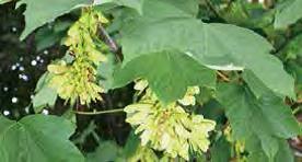
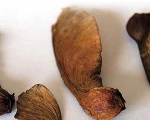
Atypical myopathy is a painful and potentially fatal muscle disease caused by a toxin found in the seeds of trees of the genus Acer, for example the sycamore tree and box elder tree. The disease is most commonly seen at this time of the year so it is important to take steps to prevent it!
Horses that develop AM are often kept on sparse pastures containing an accumulation of dead leaves, wood, and trees, and these animals often are not fed any supplementary hay or feed. The seeds are not palatable for horses but animals grazing on poor quality pasture may ingest sufficient amounts to result in signs of disease.
Prevention:
Horse owners are advised to identify these trees on or near grazing land and take steps to prevent the seeds falling where they are in reach of horses, including:
- Restrict access to seeds by using temporary fencing
- Ensure horses have access to good quality uncontaminated pasture
- Move horses off pasture at times of risk
- Provide supplementary feed in the field to minimize the risk of horses being tempted to ingest seeds
- Be aware that a field without sycamore trees can still contain seeds spread by high winds or flood waters
- Monitor horses closely to pick up any signs of a problem in the early stages
Laryngeal Ultrasound in Yearling Thoroughbreds with Two Year Old Follow Up – 2019
April 21, 2020
Laryngeal Ultrasound in Yearling Thoroughbreds with Two Year Old Follow Up
B Hunter DVM MS MANZCVS DACVS-LA
D P Keenan BVSc MANZCVS
A Ritmeester BVSc (Hons) MS DACVS
D Hanlon BVMS MVSc PhD DACT
Reason for performing study: Echogenicity of the left cricoarytenoideus lateralis muscle (CALM) has been reported to be 90% sensitive and 98% specific for diagnosing left recurrent laryngeal neuropathy in racing Thoroughbreds ≥ 2 years of age (1), but assessment of this same muscle in yearlings has not been reported.
Objectives: To determine the echogenicity of both the left and right CALM in clinically normal yearling Thoroughbreds intended for racing and correlate echogenicity with grade of arytenoid function on standing endoscopy. To repeat ultrasound and endoscopy assessments in the same population as two year olds. Also, to determine the incidence of laryngeal cartilage anatomic abnormality and correlate with endoscopic abnormality.
Hypothesis: As left arytenoid function on endoscopy decreases, echogenicity of the left CALM will increase.
Methods: Thoroughbreds born in 2014 and 2015 underwent standing endoscopy when 12-14 months of age and left laryngeal function was graded 1-5 using the Lane system. Following endoscopy, horses were sedated with xylazine (0.3-0.4 mg/kg) and standard, previously described (2) transverse and longitudinal views of the CALM and associated laryngeal cartilages were acquired with a Sonoscape S2V ultrasound unit using a linear probe at 6-7 MHz and a depth of 5.5 cm. All images were collected by one individual blinded to endoscopy grade (B. Hunter). All endoscopies were performed by one individual with an Olympus uniocular 1m endoscope (D. Keenan). Echogenicity of the CALM was compared to the ipsilateral thyrohyoid muscle and subjectively judged as normal or increased. The arytenoid, thyroid and cricoid cartilages were described as normal or abnormal. Still images were assessed by two reviewers blinded to horse age and endoscopy findings (A. Ritmeester and D. Hanlon). Multivariable statistical analyses were used to determine the effect of age and gender on left laryngeal function and CALM echogenicity. All analyses were performed using R version 3.3.2. Means are presented as mean ± SD. Significance was determined at P < 0.05.
Results: Endoscopy grade incidence in yearlings (n = 254) was 54.3% grade 1, 37.4% grade 2, 8.3% grade 3. There were no yearlings with a grade of 4 or 5. On re-assessment as two year olds (n = 53), incidence was 75.5% grade 1, 13.2% grade 2, 11.3% grade 3. There were no two year olds with a grade of 4 or 5. The mean age of yearlings at the time of endoscopy (1.2 ± 0.13 years) was not significantly different from the mean age of yearlings at the time of ultrasound (1.2 ± 0.12 years). Mean two year old age at endoscopy (2.3 ± 0.17 years) was not significantly different from mean two year old age at ultrasound (2.3 ± 0.18 years). Yearlings comprised 139 colts and 115 fillies. Two year olds comprised 8 colts and 45 fillies. Gender was not significantly associated with endoscopy or ultrasound findings. Twenty-nine two year olds showed no change from yearling endoscopy grade. Seventeen improved in grade and seven degenerated in grade. CALM echogenicity was not significantly associated with endoscopy grade for either observer. Reviewer one found significantly more ultrasound images to be non-diagnostic than reviewer two (P<0.05). Reviewer two found significantly more yearlings to have increased left CALM echogenicity than reviewer one (P<0.05), however only two of these had increased echogenicity as two year olds. Both of these horses were endoscopy grade 2 or less as yearlings and two year olds. Three horses had an abnormally shortened right thyroid cartilage on ultrasound with only one having an endoscopic abnormality (rostral displacement of the palatopharyngeal arch), making an incidence of 1.2% of laryngeal dysplasia in a clinically normal yearling Thoroughbred population and a 0.4% incidence in endoscopically normal yearling Thoroughbreds.
Discussion: Left CALM echogenicity does not correlate with endoscopy grade in clinically normal yearling Thoroughbreds that are grade 3 or less using the Lane grading system. Although all images were collected by one individual, subjective assessment of CALM echogenicity on still images has high inter-observer variability. Thus, laryngeal ultrasound is not reliable as a screening tool for subclinical left recurrent laryngeal neuropathy in clinically normal yearlings but can be used to screen for laryngeal dysplasia. The two year old gender distribution had a disproportionately high number of fillies, as colts are more commonly sold at yearling sales while fillies are kept home to race in this population.
References:
- Garrett KS, Woodie JB, Embertson RM. Association of treadmill upper airway endoscopic evaluation with results of ultrasonography and resting upper airway endoscopic evaluation. Equine Vet J 2011;43:365-371.
- Garrett KS, Embertson RM, Woodie JB, Cheetham J. Ultrasound features of arytenoid chondritis in Thoroughbred horses. Equine Vet J 2013;45:598-603.
SAA in horses: a useful screening tool for infections – May 2017
May 15, 2017
SAA in horses: a useful screening tool for infections
Cordelia Francis BVSc MRCVS
Serum Amyloid A (SAA) is an “acute phase protein”, which is a collective term for proteins that are produced and released by the liver in response to inflammation. It is a useful marker for several reasons. Firstly SAA levels increase very rapidly in response to infection, often within 24 hours, which is helpful for making an early diagnosis. Secondly there is a huge pathologic range; normal horses have blood SAA concentrations very close to 0 mg/L and with inflammation it can rise to >3000 mg/L. This large range allows us to “grade” the severity of inflammation and can also be used to monitor the horse’s response to treatment.
SAA measurements can also be helpful in differentiating between diseases that have similar clinical signs but very different treatments. For example, a painful, swollen joint can be due to either a traumatic synovitis or a septic arthritis. Traumatic synovitis is inflammation of the joint due to injury but without infection, similar to a joint sprain in humans. Septic arthritis is inflammation of the joint caused by infection (i.e. the presence of bacteria). In the early stages these two diseases can look very similar but we can use the horse’s SAA levels to help distinguish between them. We can measure SAA levels in either blood or joint fluid samples. When there is an infection present the SAA levels are usually dramatically increased, where as they only increase mildly in traumatic synovitis. This can help us make an early diagnosis, allowing appropriate treatment and thereby improving the prognosis.
We routinely measure blood SAA levels in sick horses as any generalized infection will cause an increase, not just joint infections. Because the SAA kit we use at MVS is portable we are able to measure SAA levels both at the clinic and on the farm.
Update on tetanus prevention – April 2017
April 23, 2017
Update on tetanus prevention – April 2017
Marcus Allan, BVSc
In a recent vet meeting at the Matamata Veterinary Services Equine Hospital we discussed tetanus, with an emphasis on appropriate preventative recommendations based on current scientific literature.
Tetanus is caused by the bacterium Clostridium tetani, which forms spores that survive in soil. The spores are widespread in the environment and very resistant to being killed. The bacterial spores gain access to the horse’s body through wounds that are contaminated by soil, such as deep puncture wounds, foot abscesses, surgical wounds, the umbilicus in foals and the reproductive tract in mares post-foaling. The bacterium is anaerobic and favours an environment with low oxygen levels such as that found in bruised or traumatised tissues. If conditions are favourable for growth the spores are activated and the bacteria start to multiply. The incubation period of the disease can be several weeks and therefore the original wound may be healed by the time clinical signs of tetanus develop.
- tetani produces toxins that bind to the nervous system, resulting in the typical clinical signs of tetanus. These include an exaggerated response to stimuli (hyperaesthesia), rigid posture (“saw horse” stance), stiff gait, elevated tail, difficulty opening the mouth (“lockjaw”), inability to eat (dysphagia), altered facial expression (lips pulled back into grimace, flared nostrils, erect ears, prolapsing or flicking the third eyelid across the eye), generalised muscle spasms, inability to walk, falling over, “star-gazing” (opisthotonus) when lying down and seizures, all of which quickly progress in severity. Affected horses usually die from respiratory failure, or are subjected to euthanasia.
The prognosis for surviving clinical diseased caused by C. tetani is very poor, with 60-85% of affected horses dying or being euthanased. Therefore, disease prevention through vaccination is strongly recommended. However, recommended vaccination protocols vary significantly between countries, prompting our review of the current literature.
There was much discussion about the use of tetanus vaccine (toxoid) versus the anti-toxin. The toxoid stimulates long-lasting immunity but takes at least 2 weeks to start producing immunity and around 6 weeks before strong immunity develops, whereas the anti-toxin provides immediate protection against tetanus that lasts approximately 3 weeks before wearing off. One potential concern with use of the anti-toxin is the possibility of inducing serum hepatitis. This is an immune reaction of the recipient horse to the blood proteins of the donor horse used to create the anti-toxin. The chance of this happening is very low but when it occurs it can be a serious complication. For this reason we prefer to use the toxoid alone whenever possible prior to surgery or when treating a wound; however, if the horse’s vaccination status is unknown it is essential to give the anti-toxin as well to provide that immediate protection. Because the toxoid takes at least 2 weeks to produce some degree of immunity, if the horse is unvaccinated the tetanus bacteria could invade and start producing toxins before the horse’s immune system becomes activated by the toxoid.
Given these facts, Matamata Veterinary Services now make the following recommendations:
- Unvaccinated horses (or vaccination status unknown)
- 2 vaccinations 4 weeks apart, followed by a booster dose 1 year later
- Foals start vaccinations at 3 months of age or older e.g. at weaning
- Vaccinated horses (maintenance)
- Booster dose every 3-5 years
- Horses with wounds (or having surgery)
- If vaccinated within the previous 3-5 years then boost with vaccine
- If vaccinated >3-5 years ago (or vaccination status unknown) then boost with vaccine and give anti-toxin simultaneously
- This should be followed up with a second vaccine at 4 weeks and a booster dose 1 year later for full protection
- Pregnant mares
- If vaccinated within the previous 3-5 years then boost with vaccine 4-6 weeks before foaling
- If vaccinated >3-5 years ago (or vaccination status unknown) then vaccinate 8-10 weeks before foaling so second dose is given 4-6 weeks before foaling
- New-born foals
- Give anti-toxin at birth if mare was not vaccinated during pregnancy (or her vaccination status is unknown) or if foal has not received adequate colostrum (regardless of mare’s vaccination status)
Keeping accurate tetanus vaccination records is the best way to ensure appropriate tetanus prevention is given when required. This is not always easy due to horses being moved and sold frequently. Vaccination status should be discussed before horses are moved or sold so the new manager or owner is well-informed and able to make the correct decisions to protect the horse from this devastating disease.
Gastric Ulcers – How do I know if my horse’s stomach hurts? – October 2016
October 10, 2016
Gastric Ulcers – How do I know if my horse’s stomach hurts? – October 2016
Barbara Hunter, DVM MS DipACVS-LA
Registered Specialist in Equine Surgery
Gastric ulcers are a common problem that plague both humans and horses, and any of you that have had a stomach ulcer will fully sympathize with the burn in an affected horse’s gut. The disease process is slightly different between humans and horses, due in part to the anatomic variation in the stomach lining that horses have. This article will briefly explain the gastric ulcer disease process in horses, the incidence of ulcers in various breeds and athletic types, how to diagnose ulcers, treatment recommendations and suggestions to minimize occurrence.
Equine gastric ulcer syndrome is broken into two different syndromes based on the location of the ulcer in the stomach; equine squamous gastric disease (ESGD) and equine glandular gastric disease (EGGD). The horse stomach is has an upper and a lower portion, each having a different type of lining (Figure 1). The upper portion is lined with the same lining that is in the esophagus. This is a tough, squamous cell lining that produces minimal secretions and is resistant to damage from contact with roughage. It has limited contact with stomach acid in the normal horse and develops ulcers when in excessive contact with acid. The lower portion of stomach wall has a soft, glandular lining. This region is in contact with digestive juices nearly continuously and produces most of the stomach secretions, including the HCl acid responsible for initial digestion of feed.
In the horse, equine squamous gastric disease is the considerably more common form of gastric ulceration and is also the form that is better understood both for pathophysiology and for treatment. They occur more commonly in training horses and in horses that have empty stomachs throughout the day. In both cases, increased exposure of the squamous portion of the stomach to acid precipitates ulcers. Equine glandular gastric disease is less common. Its causes are poorly understood and response to treatment is unpredictable.
Equine squamous gastric disease can be broken into two forms. Primary disease is seen with intensive feeding, environmental management and intense athletic performance but the gastrointestinal tract is otherwise normal. Secondary disease occurs due to underlying disease such as delayed gastric emptying (as one sees with an abnormally narrow exit to the stomach) or a stomach impaction. Primary ESGD is by far the more common syndrome with Thoroughbreds being most commonly affected. Studies have shown that nearly 37% of untrained Thoroughbreds have ESGD, and 80% or more develop ESGD within 2-3 months of entering race training. Incidence in Standardbred race horses is similar. By contrast, 17-58% of show horses have ESGD and 66% or more of endurance horses are affected with incidence increasing in more elite horses. Interestingly, additional studies have shown that Thoroughbreds seem to have an increased incidence of ESGD regardless of age and exercise level in comparison to other breeds.
With such a high incidence of ESGD in horses, how can you know if your horse is among the suffering population?? For years people have treated their horses for presumed ESGD based on clinical signs. Some clinical signs that can be associated with gastric ulceration include colic (especially after eating grain), inappetence, poor body condition, poor hair coat, weight loss, and behavioral changes such as teeth grinding, ‘girthiness’, and ‘cribbing’.
Perhaps the most disconcerting clinical sign in athletic horses, particularly Thoroughbred racehorses, is poor performance. Just the presence of ulcers seems to be able to precipitate poor performance in racehorses, regardless of size and severity.
With such a mixed bag of potential clinical signs, how can you know for sure if your horse has gastric ulcers? A definitive diagnosis can be made on gastroscopy. Similar to upper airway endoscopy, gastroscopy involves passing a flexible fiber optic endoscope up the horse’s nose. It is passed beyond the throat, down the esophagus into the stomach. Why is it advisable to get a definitive diagnosis of gastric ulcers? Three reasons: 1) If a horse does not have gastric ulcers and he is treated presumptively, money on treatment has been wasted. 2) If a horse does have gastric ulcers and is treated presumptively, if the horse’s clinical abnormality does not improve with treatment, you do not know if the treatment is failing or he just did not have ulcers. The recommended course of therapy for gastric ulcers in the squamous portion of the stomach is 28 days of daily omeprazole (Ulcershield) at the full dose. About 80% of horses will completely resolve ulcers with this treatment. That means 20% do not completely resolve. This is potentially problematic as poor performance can be only clinical sign of ulcers in some horses and this can continue regardless of ulcer severity. 3) Glandular gastric ulcers, although less common, have been reported in 44% or more of Thoroughbred racehorses. Awareness of this type of ulcers is of value when considering treatment as they typically require 2-3 months of treatment and may require medication other that omeprazole to resolve.
Treatment of ESGD is relatively straight-forward. Pre and post treatment gastroscopy is recommended to determine ulcer presence and severity initially and verify resolution prior to termination of treatment. Prevention of gastric ulcers, or more correctly, minimizing the risk for development of gastric ulcers, can be accomplished through several means. Maintaining horses in training on a ‘preventative dose’ of omeprazole (1/4 of the treatment dose) has been shown to lessen ulcer development. Additional ways to decrease risk are to provide constant access to water, continuous access to good quality grass pasture or free choice hay, and minimize grain and concentrates. Of course, this type of management is often not possible in the high performance athlete during training and competition, thus some owners opt to maintain the preventative dose of omeprazole during training with intermittent gastroscopies to monitor for ulcer development. Regardless of your approach, it is valuable to be aware that gastric ulcers are a common problem in our equine athletes and while treatment is usually straight forward and effective, that is not always the case.
Reference:
Sykes BW. et al. 2015. European college of equine internal medicine consensus statement – Equine gastric ulcer syndrome in adult horses. Journal of Veterinary Internal Medicine. 29:1288-1299.

Two lower airway respiratory problems that can decrease athletic performance – April 2016
June 1, 2016
Two lower airway respiratory problems that can decrease athletic performance – April 2016
Barbara Hunter, DVM, MS, DACVS-LA
Registered Specialist in Equine Surgery
The last article in our short series on performance limiting respiratory problems will focus on two diseases that affect of the lower airways of the horse. These diseases are known by the scientific names of ‘Inflammatory Airway Disease’ (IAD) and ‘Recurrent Airway Obstruction’ (RAO), also known as ‘heaves’. As both diseases tend to produce very similar clinical signs in their earlier stages, without the assistance of differentiating diagnostics, they are often both presumed to be the horse version of ‘allergies’ when in seen in their mildest forms. Diagnostic sampling of fluid from the mid to lower airways is necessary to differentiate between the two, help direct treatment and prognosticate likelihood of response to treatment.
Let’s start by comparing the major clinical differences between IAD and RAO. Recurrent airway obstruction tends to occur in older horses (average age of onset is 9-12 years old) while IAD can occur in any age horse (although most commonly horses are <5 years old). Both diseases are characterized by poor performance, exercise intolerance and coughing. IAD horses may or may not have excess tracheal mucous and affected horses typically have little or no clinical signs at rest. In contrast, horses with RAO tend to have increased respiratory effort at rest (hence the development of a ‘heave’ line), produce excess mucous when coughing and have nasal discharge. Horses with severe RAO may also have difficulty maintaining their weight and may show some degree of decreased appetite.
Recurrent airway obstruction results when allergens such as dust or pollen incite inflammation within the walls of the airway (bronchioles, bronchi). This inflammation causes thickening of the airway walls so that the space available for airflow becomes narrower. There are two forms of RAO; summer pasture induced and barn induced. The most important factor in treating RAO is removing the inflammatory allergen from the horse’s environment. For barn induced RAO, this is relatively straight forward as the most common allergens are dust and mold from hay. The two most effective ways to treat horses with barn induced RAO are to wet down their hay and either minimize dust in the barn (ie: wet down aisles prior to sweeping, sweep rather than use dust blower to clean aisles, feed hay from low feeders, etc) or completely remove affected horses from the barn. Horses that have pasture induced RAO are more difficult to treat. Moving them into a barn can sometimes improve them, but often does not. Sometimes these horses must be moved to a different geographic region before they improve.
Inflammatory airway disease, although it can occur in horses of any age, is more typically seen in young, athletic animals. Specifically, this is a disease of young racing Thoroughbreds. If you were to look at the walls of the airways on endoscopy in horses with IAD and RAO, you would be hard pressed to tell the two diseases apart as both cause airway inflammation. Differentiation between the two diseases is made on cytology of the airway fluid. This is where a sample of the fluid from the lungs is taken (the most common collection procedure is called a BAL or BronchoAlveolar Lavage) and the cells within that fluid are analyzed. Horses with RAO will have high counts of the inflammatory cells known as neutrophils while horses with IAD will have moderate amounts of neutrophils and increased numbers of mast cells and eosinophils (two other types of inflammatory cells). The importance of differentiating between the two diseases may seem academic on the surface, but in truth there can be quite significant differences in prognosis with each disease. Treatment for both diseases tends to be similar as a first line of defense: decrease exposure to environmental allergens and start on systemic corticosteroids. Horses with RAO can often be managed very well by controlling their environment after their airway inflammation has been toned down with an initial course of steroids. That is less likely to be the case in horses with IAD. Due to the presence of mast cells, IAD has more of a tendency to be refractory to treatment. Horses often need to undergo a period of rest in addition to treatment with corticosteroids to get control of airway inflammation. Gaining control of inflammation can be more difficult (longer courses of steroids, sometimes inhalant steroids are needed) and once the horse goes back into hard work, inflammation returns and the cycle starts again. Ultimately, severe cases of IAD can be just as performance limiting as grade 4 or 5 ‘roarers’.
In summary, if you are looking at an off the track Thoroughbred that is coughing (during exercise or at rest), has exercise intolerance and/or has nasal discharge, it may be prudent to look a little further into the matter. It could be that the horse just has a cold, or it could have some degree of RAO or (more commonly) IAD. Knowing whether one of these diseases is present, and to what degree, will help you determine if your horse will benefit from specific management and it will also help you determine if the horse has fit lung capacity for his or her new job.
Two upper respiratory problems that can result in horses being ‘gone in the wind’
July 26, 2015
BTB Roarers July 2015
Two upper respiratory problems that can result in horses being ‘gone in the wind’
Barbara Hunter, DVM, MS, DACVS-LA
Registered Specialist in Equine Surgery
Last month’s article focused on ‘roaring’, the most common cause of being ‘gone in the wind’ that we see in Thoroughbred horses. This month, I will talk about two more upper respiratory problems. Although epiglottic entrapment and arytenoid chondritis are not as common as roaring, both can cause abnormal noise during exercise and exercise intolerance. Thus, they can be included in the group of problems that result in wind issues in Thoroughbreds.
Let’s start with epiglottic entrapment. The epiglottis is the triangular cartilaginous structure that sits on the floor of the airway, just in front of the larynx (Figure 1a). Its job is to flip backwards and seal over the airway each time the horse swallows food, thereby preventing aspiration of the food bolus. Like most anatomic structures, there is quite a bit of variation between horses. Researchers have established that the normal length of the Thoroughbred epiglottis is 8.5 – 8.76 cm from base to tip (as measured on radiographs). The epiglottis is normally slightly arched and has plump, scalloped edges. Some horses have what is known as a hypoplastic, flaccid epiglottis (Figure 1b). This type of epiglottis is abnormally short (6.5 – 7.5 cm), and can be thin and flat with edges that curl in. In comparison to a normal epiglottis on upper airway endoscopy, this epiglottis looks short and weak. While any epiglottis can become entrapped, this is the type of epiglottis that is more prone to entrapment.
The entrapment is a result of the thin mucosal tissue underneath the epiglottis slipping up and over the epiglottis. Not only does the entrapment reduce the normal function of the epiglottis, each time the horse breathes out it breathes pressurized air into this film of tissue that billows up and slows outward airflow. As the problem becomes more chronic, this tissue becomes thickened, ulcerated and painful. The clinical signs associated with entrapment in a racehorse include a whistling noise during exercise and decreased performance. In sport horses, the more common clinical sign is coughing, particularly during collection. Treatment is straight forward. The entrapping membrane is cut via a quick and simple procedure (Figure 2) and the horse is placed on a course of anti-inflammatory throat wash combined with systemic anti-inflammatories (eg: bute) and antibiotics (typically Sulpha-T). Prognosis for return to function is good with most horses returning to full function within 3-6 weeks of initial treatment.
Arytenoid chondritis is an infection of the arytenoids (Figure 3) that can affect horses of all ages and disciplines. We even see this problem in broodmares turned out to pasture. Clinical presentation is typically heard as abnormal noise on both inspiration and expiration. Most commonly the problem is trauma induced. Basically the left and right arytenoids slam together during periods of upper airway activity (excessive whinnying, heavy breathing associated with extreme athletic activity) and the trauma associated with this motion results in damage to the mucosal surface of the arytenoids. The bruised, inflamed arytenoid mucosa then becomes mildly infected with the horse’s own natural upper respiratory tract bacteria and this infection prevents healing and encourages more inflammation. Diagnosis is made with upper airway endoscopy. Mild cases tend to be associated with superficial mucosal lesions and often these types of lesions have no outward clinical signs. These are the types of lesions that are seen in yearling Thoroughbreds on endoscopy at Sales and are typically secondary to them calling out excessively in the exciting Sales environment. The lesions are treated with an anti-inflammatory throat wash ± systemic antibiotics depending on severity.
A more severe form of arytenoid chondritis is the formation of granulomas on one or both of the arytenoids (Figure 3). These are literally little balls of granulation tissue that protrude into the airway, obstructing airflow and causing respiratory noise. In severe cases they can become large enough to completely cut off air supply, but this is relatively uncommon. Treatment for these is also relatively straight forward. Most small granulomas respond well to systemic antibiotics and anti-inflammatories combined with an anti-inflammatory throat wash. Larger granulomas should receive the same treatment, but the granulomas may need to be resected in addition to those treatments.
Severe cases of arytenoid chondritis can infect the underlying cartilage as well as the mucosa. This is a less than ideal situation as infected cartilage is quite difficult to resolve with antibiotics. Often these horses require either a protracted course of antibiotics and throat wash, or surgical resection of the affected arytenoid. In some animals, even if the infection of the cartilage gets cleaned up with antibiotics, the infection can cause distortion of the cartilage, which can in turn obstruct the airway and limit athletic performance. As a result, those horses also need surgical resection of the affected arytenoid in order to return to full racing function, but exercise intolerance is typically not seen in less aerobically demanding sports. Given the knowledge that the cost of treating and prognosis associated with infected cartilage versus more superficial chondritis is quite different, I do recommend determining if cartilage is involved in a chondritis if the chondritis does not resolve with a standard 2 week course of medical therapy. Laryngeal ultrasound is a relatively new technique that takes some experience to perform, but cartilage involvement can be fairly easy to determine with ultrasound in the hands of an experienced practitioner (Figure 4a and b).
In summary, epiglottic entrapment and arytenoid chondritis are two additional upper respiratory abnormalities that can cause a horse to present clinically as ‘gone in the wind’. Both are easily diagnosed with upper airway endoscopy. Most cases are treatable with good results. Laryngeal ultrasound is recommended for arytenoid chondritis cases that do not respond appropriately to treatment as determination of cartilage involvement can affect prognosis and treatment duration.
Roaring, epiglottic entrapment and arytenoid chondritis are three of the most common upper respiratory tract problems that can cause a horse to be ‘gone in the wind’. The article for next month will be the last of our short series on respiratory issues and will take a look at lower airway problems (eg: heaves, inflammatory airway disease) that can cause decreased athletic performance.

Figure 1a: Normal Epiglottis

Figure 1b: Hypoplastic epiglottis
(Garrett et al. 2010)

Figure 2: Cutting the membrane that causes an entrapped epiglottis (Lacourt et al. 2011)

Figure 3: Arytenoid chondritis with large granuloma (photo courtesy of Dr. A. Ritmeester)

4a: Longitudinal view of a horse with a normal left arytenoid and a severely thickened, misshaped right arytenoid with right sided arytenoid chondritis.

4b: Transverse view of the horse in 4a. Note the severely thickened right arytenoid cartilage in comparison to the normal left.
References:
Garrett KS, Pierce SW, Embertson RM, Stromberg AJ. Endoscopic evaluation of arytenoid function and epiglottic structure in Thoroughbred yearlings and association with racing performance at two to four years of age: 2,954 cases (1998-2001). JAVMA 2010;236:669-673.
Lacourt M, Marcoux M. Treatment of epiglottic entrapment by transnasal axial division in standing sedated horses using a shielded hook bistoury. Vet Surg 2011;40:299-304.
BTB Roarers June 2015
July 5, 2015
BTB Roarers June 2015
I’m considering an ‘off the track’ Thoroughbred as my next riding horse, but I hear the one I like is ‘gone in the wind’. Should I look elsewhere for what I need?
Barbara Hunter, DVM, MS, DACVS-LA
Registered Specialist in Equine Surgery
The first step in answering this question is to describe what is meant by the term ‘gone in the wind’. The second is to define what you need your retired racehorse to do in his or her new job.
The term ‘gone in the wind’ is a relatively loose term that is used to describe a multitude of upper airway abnormalities in the horse that result in respiratory noise during exercise. By far the most common upper airway abnormality that it is used to describe in Thoroughbreds is a syndrome called recurrent laryngeal neuropathy (RLN), also known as ‘roaring’. This is a syndrome where degeneration of the recurrent laryngeal nerve results in loss of the muscles that control movement of the left arytenoid (the left and right arytenoids are the cartilage flaps that close the entrance of the windpipe during swallowing to protect against aspiration of food [Figure 1]). The result of this muscle loss is that the left arytenoid cartilage literally ‘flaps in the breeze’ each time the horse breathes in. Horses affected with RLN are commonly called “roarers”, due to the characteristic roaring noise these horses make on inspiration during exercise. The focus of this article will be on roarers, but the term ‘gone in the wind’ can refer to respiratory issues other than roaring such as epiglottic entrapment , arytenoid chondritis, dorsal displacement of the soft palate or recurrent airway obstruction (aka: heaves). Although all of these problems have potential to cause noise and variable degrees of exercise intolerance, treatment for each of them is different and the prognosis associated with treatment can vary. In an effort to keep these articles relatively succinct, I will address these problems separately in future articles over the next few months. Suffice to say, however, before deciding whether a ‘gone in the wind’ Thoroughbred will meet your needs, it is essential to obtain an accurate diagnosis of what the actual problem is.
Let’s start by discussing how to accurately diagnose a roarer. Although the sound a roarer makes is fairly characteristic, unfortunately it is by no means a 100% accurate means of diagnosing the problem. More importantly, the degree of noise made is at best a rough estimate of the severity of the problem. An accurate diagnosis is best made via upper airway endoscopy. This can be done at rest (resting endoscopy) or during exercise (dynamic endoscopy). Laryngeal ultrasound is also a useful adjunct diagnostic tool. Dynamic endoscopy is considered the gold standard diagnostic for almost all upper airway abnormalities in the horse. In the case of roarers, moderate to severally affected horses will tend to show the abnormality on resting endoscopy, while mildly affected horses often need the strain of a hard gallop to induce the abnormality. With this in mind, you can already surmise that if you are not planning to ask your ‘off the track’ Thoroughbred to do a sport that involves hard galloping, a mild case of RLN is fairly unlikely to cause exercise intolerance.
That begs the question then, how do we differentiate a mild roarer from a severe one. The answer: we grade them. There are two grading systems that veterinarians use to grade roarers and the details of these two systems are compared in Table 1. North America and Europe use the Havermeyer system of grading while Australia and New Zealand use the Lane system. Using the Lane system, a grade of 4 or 5 is expected to negatively affect racing performance in a Thoroughbred. It is important to remember though that racing puts more strain on the upper respiratory tract than any other sport. Thus, while a moderate roarer may be a severe problem for a race horse, it is unlikely to cause performance limiting exercise intolerance in a pleasure riding horse, dressage horse or low level jumper.
On the converse side, if you are looking for an intermediate level eventer and your perfect Thoroughbred is a grade 5 roarer, this horse is likely to need some help in order to meet your needs. The ‘tie-back’ and ventriculo-cordectomy are the combination of surgical procedures commonly used in roarers to stabilize their upper airway. The ‘tie-back’ uses high tensile suture to literally tie the dysfunctional left arytenoid to the side of the larynx so that it is not flapping freely in the middle of the airway with each breathe. This is the portion of the procedure that is needed to improve exercise tolerance in galloping horses. The ventriculocordectomy is a procedure in which a small portion of the vocal cord and the little air sack (ventricle) immediately adjacent to it is removed. These two structures are part of the mechanism that help a horse to whinny. In a roarer, however, they become stretched and floppy so they can impede airflow to some degree during intense exercise. These are also the structures that cause most of the abnormal noise that a roarer makes. In a dressage horse this is an important consideration as dressage horses can be eliminated from competition due to ‘wind unsoundness’. As the aerobic demands of a dressage horse are significantly less severe than those of a racehorse, exercise intolerance secondary to roaring is not commonly seen. Thus, a ventriculo-cordectomy to resolve the noise may be all that is needed if you are considering a high grade roarer as a potential dressage horse. This differentiation is worth noting as a ventriculo-cordectomy is about a third of the cost of a tie back/ventriculo-cordectomy combination and can be done under a brief intravenous general anesthetic (similar to a castration type of anesthetic).
In summary, if you are considering an ‘off the track ‘ Thoroughbred as your next sport horse, don’t eliminate horses that have a history of being ‘gone in the wind’ from your list of candidates if they otherwise meet your criteria as an athlete. First, find out if they truly are roarers, or if ‘gone in the wind’ refers to another wind condition. Secondly, find out what grade of roarer the horse is. Horses that are grade 3 or less are still likely to make good athletes in sports that do not require them to do hard galloping. Lastly, if the horse is a grade 4 or 5 roarer, but otherwise seems like the perfect candidate for you, investing in a surgical procedure that improves his airway is always an alternative.
As mentioned earlier, ‘gone in the wind’ can refer to many different upper respiratory problems that cause noise. Next month’s article will address epiglottic entrapment and arytenoid chondritis as causes of respiratory noise and what potential these problems have to influence performance in sport horses or pleasure horses.


