


Endoscopy
Endoscopy
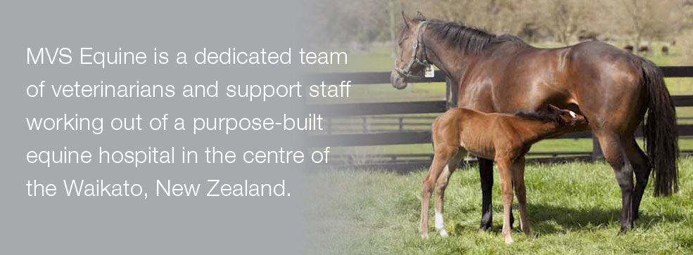
November 22, 2011
Endoscopic evaluation of a horse’s upper airway is most commonly performed at rest. Resting endoscopic examination of the upper airway is an effective diagnostic tool for identifying structural abnormalities of the larynx.
These include:
- Laryngeal hemiplegia
- Rostral displacement of
- The palatopharyngeal arch
- Epiglottic entrapment
- Lymphoid hyperplasia
- Epiglottic hypoplasia
- Subepiglottic cysts
- Arytenoid chondritis
- Persistent dorsal displacement of the soft palate
Abnormalities that decrease the diameter of the airway and increase airway resistance can cause respiratory noises due to air turbulence. This is heard as a roar or a whistle during inspiration at exercise. If the degree of obstruction is severe enough, then exercise intolerance or poor performance may result.
In marginal or subtler cases the evaluation is repeated using video endoscopy, which results in an easier procedure for the examiner. Videotapes of the examination may then be reviewed at a slower rate to accurately assess the type and degree of obstructive abnormality. Ultimately, this can be done while the horse is exercising on a treadmill to show dynamic changes that may not be seen at rest.
Laryngeal Hemiplegia … in more detail
This is a disorder whereby the symmetry in movement of the rim and vocal cords of the larynx are affected causing a functional obstruction during exercise. This is due to a partial or complete paralysis of the nerve that supplies, usually the left, vocal cord. These abnormalities in resting laryngeal function involve the corniculate process of the arytenoid cartilage (vocal cord) and have been categorised into five grades:
- Grade I: all movements are synchronised
- Grade II: all major movements are symmetrical and full abduction is achieved, although it may be delayed, especially on the left side
- Grade III: full abduction is still achieved although activity on the left side is reduced and it may appear asymmetrical at rest
- Grade IV: the left side is not capable of full abduction. Asymmetry is obvious
- Grade V: true hemiplegia. Complete paralysis and no movement on the affected (usually left) side
Grades IV and V fail the scoping panel examination and Conditions Of Sale (Section 16) at the Karaka Yearling Sales as it is likely severe enough to limit the horse’s performance.
Treatment of horses exhibiting laryngeal hemiplegia involves performing a prosthetic laryngoplasty (tie-back procedure). Prognosis for a successful return to previous level of work after laryngoplasty is reported to be 50% to 70%. Resection of the vocal cords and Hobday operation (ventriculectomy) may also be performed at the same time to help enlarge the opening (rima glotties) during exercise and this will help with eliminating the roaring noise usually heard.


Dentistry
November 21, 2011
Everybody who works with horses has an opinion about horses’ teeth. When should they be done, by whom and how often? Do you take wolf teeth out?And do you need to use sedation? There has recently been debate, at times heated, around the world about who should be doing horses’ teeth. Basically someone competent and who has undergone formal training gets our vote. There are not enough vets in the country to properly do all the horses’ teeth. Correspondingly some horses will require sedation to be done properly and some will require procedures greater than just routine rasping provided by a lot of equine dental technicians. This is where we can definitely help.
A routine dental examination and rasping is designed to serve four purposes:
- to remove sharp enamel points
- to improve mastication (chewing), which enhances the digestion of feedstuffs
- to alleviate stress on abnormally worn teeth
- to prevent discomfort associated with the bit.
A dental care programme actually starts from when a foal is born, as we inspect them at this time for any birth defects like a parrot mouth (over shot) or sow mouth (under shot), or a cleft palate. The next examination should coincide with yearling preparation at around nine months of age. This is also the time when all of the deciduous (temporary) teeth have erupted. At this time we should be removing sharp enamel points, any developing hooks, and considering removing any wolf teeth.
The horse, at this time, is also learning to accept a bit. Bits are for communication and it is not necessarily the type of bit used but rather how it is used (or misused) that can cause a problem. Trauma associated with a bit can be due to excessive pressure on the bars or soft tissue injuries to the tongue or cheeks. The cheeks are pulled back onto the edges of the first cheek teeth, especially the lowers, and if these are sharp, severe ulceration and pain will occur.
It is sensible to have a dental examination and rasp before breaking in, to reduce oral trauma and pain during this impressionable first phase of education. Bit seating is a commonly performed technique but the term is somewhat misleading, as the teeth are not actually shaped to fit the bit. In fact the bit should not be in contact with the teeth at all if it is used correctly. The concept of a bit seat is to remove the sharp prominences on the first cheek teeth that can damage the soft tissues if they are pulled hard against them. This involves essentially rounding them off. Care must be taken not to be too aggressive as it is unnecessary and may risk exposing the pulp cavity and introducing infection, especially when machine tools are used.
It is without doubt that a full mouth speculum (gag) must always be used to allow a detailed and thorough oral examination and proper routine rasping of the dental arcades. To enable this to be done safely for the horse, the handler and the person performing the examination, sedation may often be required. This needs to be assessed on a horse by horse basis. Only veterinarians may use intravenous sedatives legally in New Zealand. Wolf teeth are small (usually 1 to 2 cm) with variable size roots (0.5 to 3 cm). They erupt at between 6 to 12 months of age and can occur on the lower jaw but are more commonly on the upper jaw just in front of the first cheek tooth. Some are lost when this first temporary cheek tooth is lost at around 30 months of age. They are blamed for many behavioural problems and for interfering with the bit and are therefore frequently extracted. One argument is that they never do any good, and may sometimes do harm, so remove them. However it is also generally accepted that enlarged, abnormally placed wolf teeth are more likely to cause a problem so are good candidates for removal. Whereas normal sized and placed wolf teeth, especially in older horses showing no signs of a problem, are probably okay.
Removal is not necessarily an innocuous procedure as the roots are sometimes large, deeply embedded, and a major artery runs close by (the palatine artery). Infections and fracturing the tooth off above the gum line when attempting to remove the tooth may result in an ongoing painful local swelling.
In summary, horses up to five years of age should have their teeth checked more often (every 6 to 12 months). This is a time of rapid change in the horse’s mouth and they are going to erupt 40 teeth during this time. Serious problems can be managed or prevented with proper early intervention.
Castration
November 21, 2011
Field castration is considered to be a routine procedure that is commonly carried out. As any owner who has castrated horses knows, there is also a very high complication rate (18-33%) associated with field castration. Both owners and veterinarians have accepted this as normal. It is not uncommon to revisit a horse a week after castration to re establish drainage and administer antibiotics. Every year we also deal with a handful of chronically infected wounds requiring surgery. The cost of treating complications is often many times the cost of the original castration.
In an attempt to decrease complication rates with field castration we have recently purchased a Henderson Tool and have had great success to date. A closed castration (tunic left over the testicle) incision is used and the testicle and cord exteriorised. The tool is clamped over the cord and the other end of the tool is then placed in the drill bit. Initial turns are made to form a ‘knot’ in the cord and then the speed is increased and traction is applied. This results in a very tight seal that has very little blood loss. This decreases post castration complications including swelling, haemorrhage, trauma and infection. Due to the tight seal there is less chance for infection to enter into the abdominal cavity. This technique is faster than traditional castration and therefore decreases anaesthetic times and risks as well.
